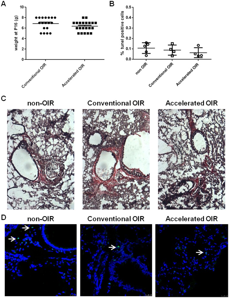
Fig 4. Histological analysis of lungs from nursing mothers after exposure to hyperoxia.
(A) Body weight of pups at P16. (B) Alveolar cell apoptosis indicated by TUNEL positive cells in lung cryosections from non-OIR control mice and mice after conventional or accelerated OIR. (C) Van Gieson staining for collagen in lung cryosections shows no fibrotic lesions in any of the groups analyzed. Original magnification: 20x. (D) CD45-staining of cryosections demonstrates few CD45-positive cells in the lungs of non-OIR control animals and those after conventional or accelerated OIR.

