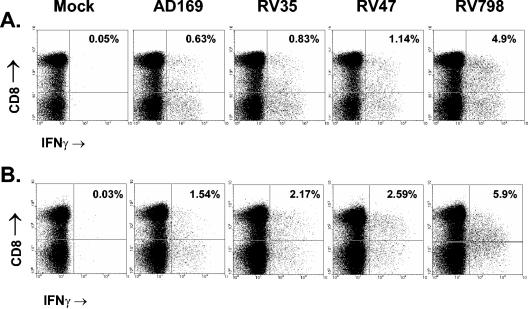
FIG. 3.
Elevated frequencies of CMV-specific T cells using recombinant CMV-infected fibroblasts. PBMC were incubated with virus-infected autologous fibroblasts for 12 h and then removed and stained with monoclonal antibodies for surface CD4 and CD8 receptors. The cells were then fixed and permeabilized, followed by staining for intracellular IFN-γ. Flow cytometric analysis was carried out, with all plots shown gated on lymphocytes by forward scatter and side scatter. Mock-infected cells were used as negative controls, and staphylococcal enterotoxin B was used to stimulate T cells as a positive control (not shown) for IFN-γ production. CD8 T-cell responses of donor 8 (A) and donor 9 (B) against mock-infected and virus-infected fibroblasts are shown.

