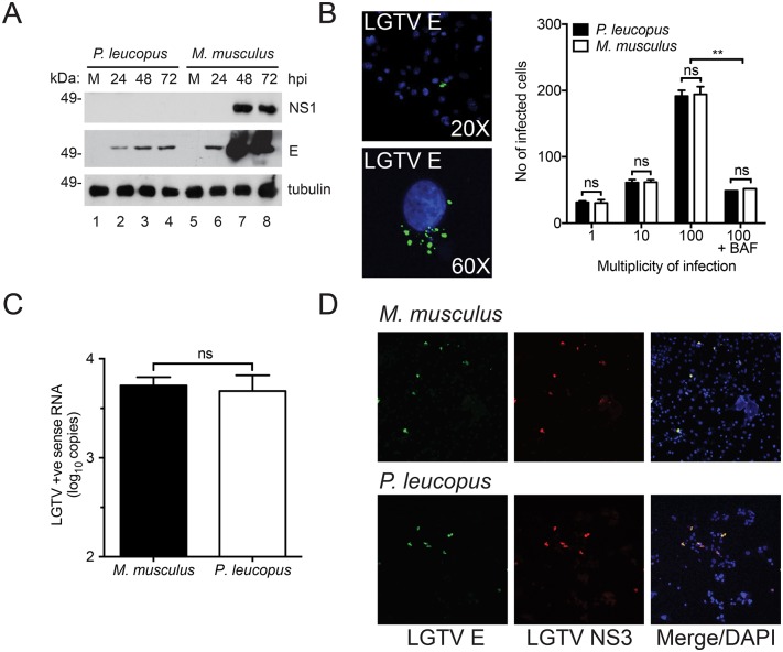
Fig 2. TBFV entry and protein expression are not inhibited in P. leucopus cells.
(A) Immunoblot of LGTV NS1 and E proteins in P. leucopus and M. musculus fibroblasts at the indicated time points post infection. (B) Representative confocal 20X (top) and 60X (bottom) images showing P. leucopus cells infected with LGTV after 1 hpi at 37°C. The viral E protein is stained in green and nuclei are stained in blue (DAPI). Quantified data are shown as the total number of infected cells at MOI 1, 10, and 100 respectively. Cells treated with bafilomycin A1 (BAF) at 15 min post-infection are shown as a negative control. (C) Viral entry assay showing abundance of LGTV positive (+ve) strand RNA in P. leucopus and M. musculus fibroblasts. The cells were infected at MOI 10 and incubated at 4°C for 1 h before a temperature shift to 37°C for another h. Total RNA was harvested following an acid wash and the resultant cDNA was used as template for RT-qPCR. (D) Confocal image showing P. leucopus and C57BL/6 expressing LGTV proteins. Cells were transfected with viral RNA for 5 days after which the cells were fixed and immunostained for LGTV E (green) and NS3 (red). Cell nuclei were stained with DAPI (blue) and visualized by confocal microscopy (20X magnification).

