Figure 1.
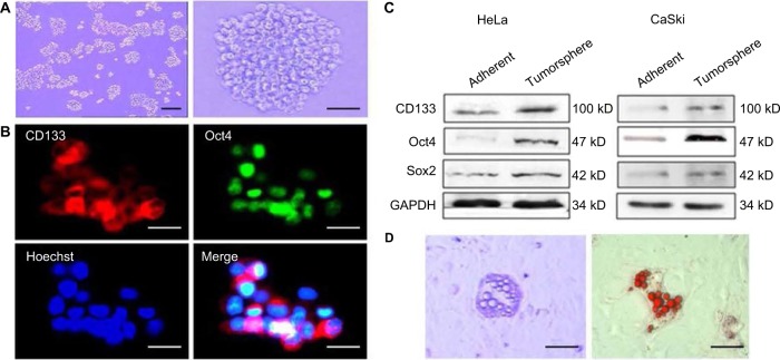
The characteristics of the cervical cancer tumorspheres. (A) Tumorsphere cultured in serum-free medium for 10–12 days. Magnification ×40 (left) and ×100 (right). (B) Immunofluorescence detected the expression of Oct4 and stem cell marker CD133 in tumor sphere cells. (C) The tumor sphere cells were further induced with lipid cells. (D) After 14–21 days of induction, the lipid drop-like oil “O” positive cells were found. Magnification ×100. Scale bars =100 μm.

