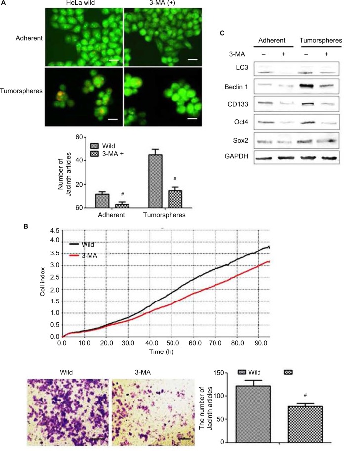
Figure 5.
The effect of autophagy inhibitor 3-methyladenine (3-MA) on cervical cancer cells. (A) Acridine orange staining detected autophagic flux in cancer stem cells and adherent cells with and without 3-MA treatment. The orange fluorescence represents autophagosomes. Magnification ×100. (B) xCELLigence RTCA (top) and Transwell (bottom) detected the effects of 3-MA on HeLa cell proliferation and invasion. (C) Western blot detected the effected of 3-MA on the level of PA proteins Oct4, Sox2 and CD133 in both adherent and tumorsphere HeLa cells. #P<0.05. Scale bar =100 μm.

