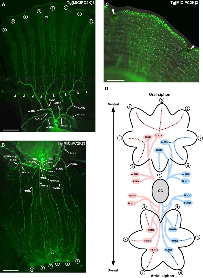
Fig 2.
(A) Innervation of the anterior nerves to the oral siphon and periphery. Anterior nerves are indicated by arrows. Arrowheads indicate tentacle row. The lobes of the oral siphons are numbered from 1 to 8. (B) Innervation of the posterior nerves to the atrial siphon and periphery. Posterior nerves are indicated by arrows. The lobes of the atrial siphons are numbered from 1 to 6. (C) Magnified image of the siphon lobe. The innervation of nerves to the edge of the siphon lobe is shown. Pigment organs are indicated by arrowheads. The small dots are Kaede-positive cells. (D) Schematic illustration of the anterior and posterior innervation to the siphons. The illustration is the overhead view of the oral and atrial siphons. The numbers of the lobes correspond to those in (A) and (B). Nerves derived from the left part of the cerebral ganglion are indicated in pink. Nerves derived from the right part of the cerebral ganglion are indicated in blue. The innervations are indicated by dotted lines. All images were taken by the fluorescence stereo microscope. AMN, anterior medial nerve; ALN, anterior lateral nerve; PMN, posterior medial nerve; PLN, posterior lateral nerve; CG, cerebral ganglion; OS, oral siphon; AS, atrial siphon. Scale bars indicate 2.5 mm in A and B and 500μm in C.

