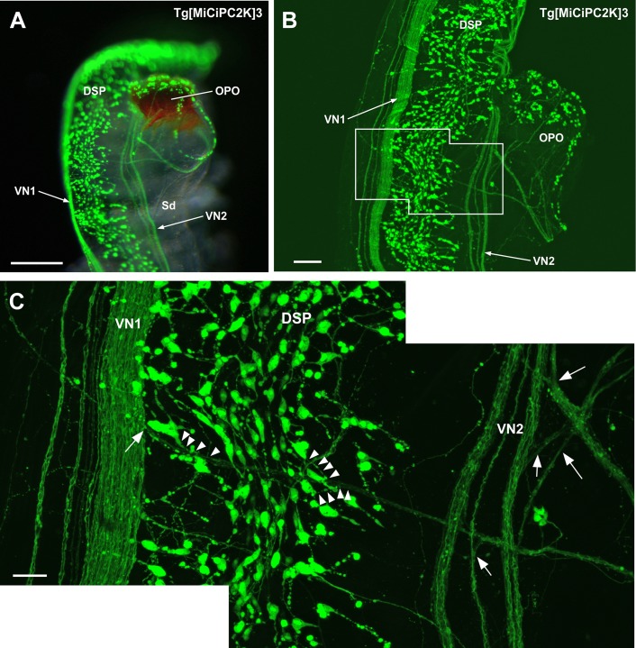
Fig 4.
(A) Image of the dorsal strand plexus, visceral nerve and orange-pigmented organ. The image was obtained by the fluorescence stereo microscope. VN1, VN2, DSP and innervation to the orange-pigmented organ (OPO) are shown. (B) Image of the dorsal strand plexus, visceral nerve and orange-pigmented organ. The image was obtained by the confocal laser scanning microscope. Dominant innervation from VN2 and minor innervation from VN1 to OPO are shown. (C) Magnified image of the framed region in (B). The branching points of the visceral nerves to the OPO are indicated by arrows. The neurons and axons innervating to the OPO are indicated by arrowheads. DSP, dorsal strand plexus; OPO, orange-pigmented organ; Sd, spermiduct; VN, visceral nerve. Scale bars indicate 250μm in (A), 100μm in (B) and 30μm in (C).

