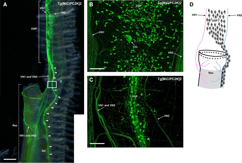
Fig 6.
(A) Image of the rectum, dorsal strand plexus and visceral nerves. The image was obtained by the fluorescence stereo microscope. The anterior part of the rectum is shown and indicated by the dotted lines. Innervations of the VN1 and VN2 to the rectum are shown. The dorsal strand plexus becomes thinner around the rectum and continues to the periphery along with the rectum and gonoduct (arrowheads). (B) Magnified image of the dorsal strand plexus above the rectum The image was obtained by the confocal laser scanning microscope. The dorsal strand plexus is large in width above the rectum and 20–30 neurons were laterally distributed. (C) Magnified image of the rectangle region in (A) The image was obtained by the confocal laser scanning microscope. The dorsal strand plexus becomes thinner and less than ten neurons were laterally distributed. (D) Schematic illustration of the innervation of the visceral nerves and dorsal strand plexus to the rectum. Neurons in the dorsal strand plexus are shown as neuron-shaped illustrations. Kaede-positive cells at the edge of the rectum are shown as open circles. VN1 and VN2 are shown in pink and blue, respectively. The region where Kaede-positive small cells are distributed is shadowed. CO, cupular organ; DSP, dorsal strand plexus; Gd, gonoduct; Rec, rectum; VN, visceral nerve. Scale bars indicate 750μm in (A) and 100μm in (B) and (C).

