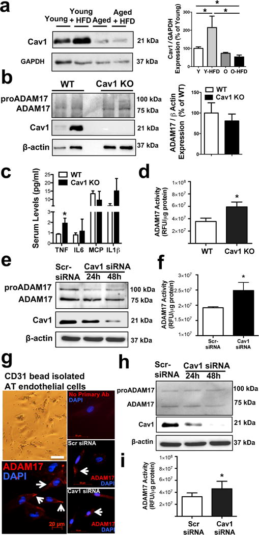
Figure 8. Loss of caveolin-1 augments ADAM17 activity in vascular endothelial cells.

Representative Western immunoblots and summary data (N=4 in each group) show Cav1 expression (normalized to GAPDH) in homogenates of adipose tissue obtained from young and aged mice with normal or HFD (panel a). Representative Western immunoblots and summary data (N=4 in each group) show Cav1 and ADAM17 expression (normalized to β-actin) in adipose tissue obtained from wild-type (WT) or Cav1 knockout mice (panel b). Serum levels of TNF, IL-6, MCP-1 and IL1β as well as adipose tissue ADAM17 enzyme activities were measured in WT and Cav1 knockout mice (panel c & d, n=4 in each group). ADAM17 and Cav1 protein expression as well as ADAM17 enzyme activities were measured in cultured human coronary artery endothelial cells (Panel e & f) or CD31 micro bead isolated and cultured human adipose tissue endothelial cell (panel g, h & i) that were transfected with scrambled siRNA control (Scr-siRNA) or with Cav1-targeted siRNAs (Santa Cruz) for 24 hours or 48 hours (ADAM17 immunocytochemistry and ADAM17 enzyme activity was assessed in cells after 48 hours of transfection, N=3 in each group). Data are shown as means ± SEM. *P<0.05.
