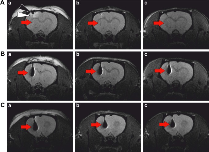
Figure 1.
MR imaging of a rat brain.
Notes: T2-weighted images after implantation of (A) unlabeled cells, (B) cells labeled by SPIONs, (C) cells labeled by CZF-NPs. Red arrows indicate the location of the cell implant. MR images were obtained (a) within 24 hours after implantation, (b) 1 week, and (c) 4 weeks after implantation. An edema (white triangle) in the left cortex was visible as a hyperintense area immediately after implantation of the unlabeled cells into the brain (A), which vanished within 1 week, whereas a small hypointense area just under the skull, originating from heme iron from blood (black triangle), remained visible until the end of the experiment.
Abbreviations: CZF-NPs, cobalt-zinc-ferrite nanoparticles; SPIONs, superparamagnetic iron oxide nanoparticles; MR, magnetic resonance.

