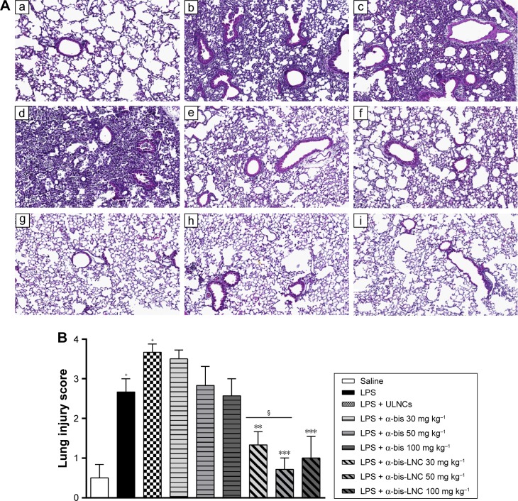
Figure 4.
Effect of treatment with α-bis-LNCs on tissue damage.
Notes: The mice were treated with α-bis, α-bis-LNCs (30, 50, or 100 mg kg−1), or ULNCs 4 hours before LPS or saline instillation. The lungs were perfused and collected in formalin. Lung serial sections were embedded in paraffin by routine methods and stained with H&E. (A) Microscopic analysis of stained tissue sections: a, saline; b, LPS; c, LPS + ULNCs; d, α-bis 30 mg kg−1; e, α-bis 50 mg kg−1, f, α-bis 100 mg kg−1; g, α-bis-LNC 30 mg kg−1; h, α-bis-LNC 50 mg kg−1; i, α-bis-LNC 100 mg kg−1; 10× magnification. (B) Histological analysis of lung inflammation by a scoring system. Data are expressed as the mean ± SEM (n=5–7). +P<0.05 compared with the saline group; **P<0.01, and ***P<0.001 compared with the LPS-induced group; §P<0.05 compared with α-bis.
Abbreviations: α-bis, α-bisabolol; α-bis-LNCs, α-bisabolol-loaded lipid-core nanocapsules; LNCs, lipid-core nanocapsules; ULNCs, drug-unloaded nanocapsules; LPS, lipopolysaccharide; SEM, standard error of mean.

