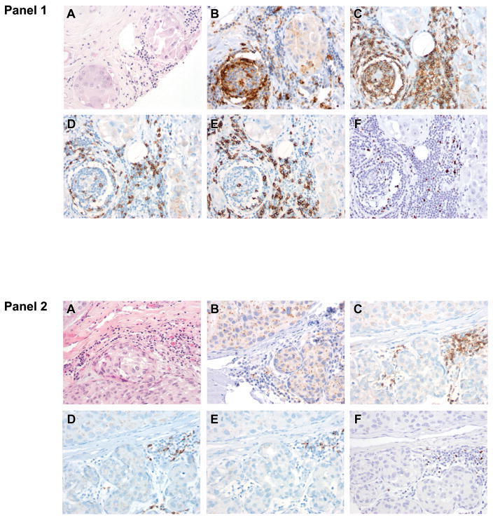
Figure 2. Immunologic features of the ductal carcinoma in situ (DCIS) tumor microenvironment.
Most DCIS cases (81%) (Panel 1; A, H&E) display PD-L1+ tumor infiltrating lymphocytes (B). PD-L1+ tumor infiltrating lymphocytes are associated with greater numbers of all tumor infiltrating lymphocyte subsets including CD4 helper T cells (C), CD8 + cytotoxic T cells (D), CD20+ B cells (E), and FoxP3 + Tregs (F) relative to DCIS with PD-L1− tumor infiltrating lymphocytes(Panel 2). Importantly, no DCIS carcinoma cell displayed cell surface PD-L1 staining .

