Figure 3.


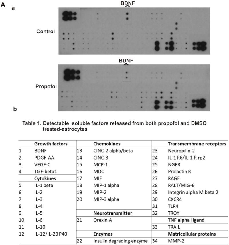
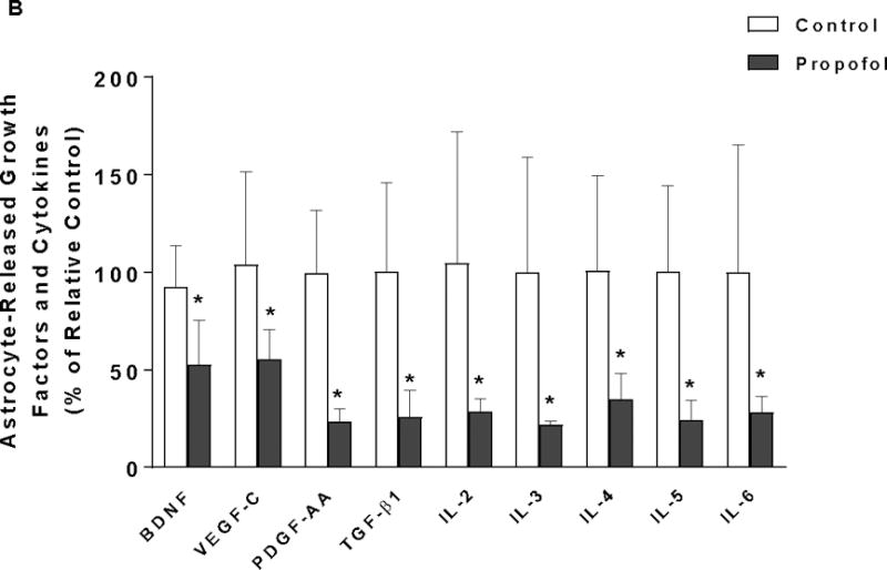
Astrocyte-derived growth factors and cytokines in conditioned medium are downregulated by propofol. (A) Analysis of paracrine factors in the conditioned medium from DMSO- or propofol-treated astrocytes using dot blot array. (A-a) The representative images of dot blot analysis of 90 soluble protein profiles in astrocyte-conditioned medium following propofol and DMSO exposure. The each double black dot indicates the individual protein that could be detected. There were 34 soluble factors detected. (A-b) The table lists the name of the 34 soluble factors [shown as black dots in (a)] that were released from both propofol- and DMSO-treated astrocytes. The protein array map refers to RayBiotech web http://www.raybiotech.com/files/manual/Antibody-Array. (B) Among 34 detected soluble factors, propofol downregulated the secretion of nine key growth factors and cytokines that are important for neuron survival [e.g., brain-derived neurotrophic factor (BDNF) was indicated in the protein array maps] from astrocytes as compared to DMSO control. Conditioned medium was collected 12 h following 6 h of 30 μM propofol exposure. The protein expression in conditioned medium was normalized as % of p-3b internal control using dot analysis. n=4. *P<0.05 vs. relative control.
