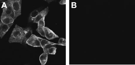
FIG. 2.
Confocal microscopic analysis of 3a protein in SCoV-infected cells. Vero E6 cells growing in eight-well chamber slides (Lab-Tek, Naperville, Ill.) were infected with SCoV at a multiplicity of infection of 1 (A) or mock infected (B). At 24 h p.i., cultures were incubated overnight with 4% paraformaldehyde and then treated with 0.25% Triton X-100 for 15 min. Subsequently, cells were incubated with anti-3a antibody and goat anti-rabbit secondary antibody conjugated with Alexa Fluor 488 dye (Molecular Probes, Eugene, Oreg.). Cells were observed under the Zeiss LSM 510 UV META laser scanning confocal microscope in the University of Texas Medical Branch Infectious Disease and Toxicology Optical Imaging Core.

