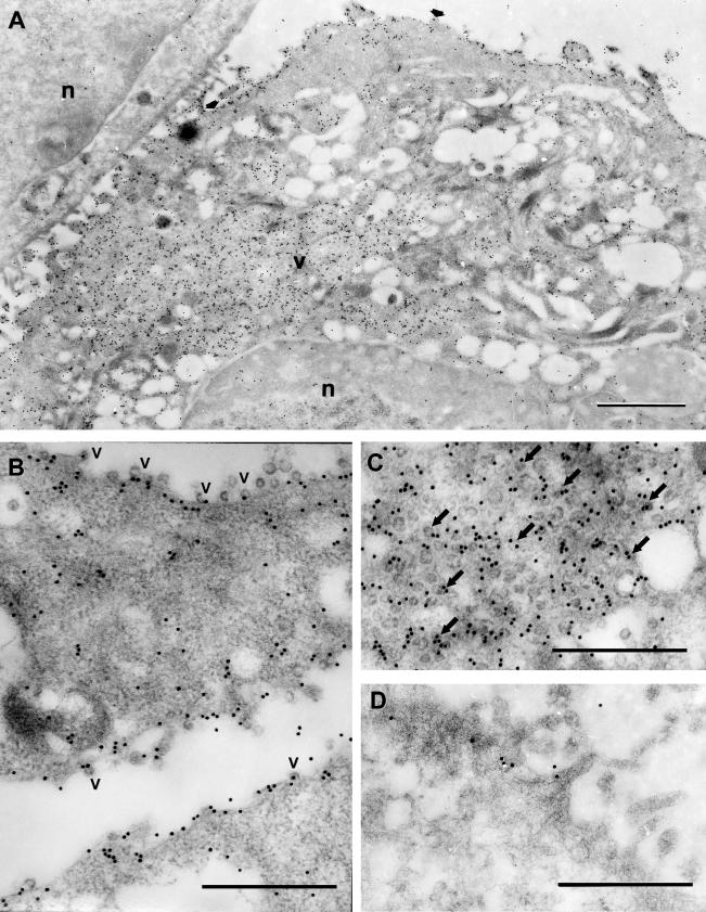
FIG. 3.
Immunogold labeling of 3a protein in SCoV-infected cells. Caco2 cells were infected with SCoV at a multiplicity of infection of 0.5, fixed at 48 h p.i., and embedded in LR White resin. Ultrathin sections of the cells were incubated with anti-3a antibody and goat anti-rabbit immunoglobulin G (heavy plus light) conjugated to 15-nm colloidal gold particles (Amersham Biosciences). (A) In a SCoV-infected cell the label is clearly associated with intracellular virus (v) and with the virions at the cell surface (arrows). An uninfected cell in the upper left corner is devoid of label. n, nucleus. Bar = 1 μm. (B) 3a protein is associated with plasma membranes. V, SCoV particles associated with 3a protein. (C) Association of 3a protein with intracellular virus particles (arrows). (D) Portion of cytoplasm of a mock-infected Caco2 cell showing occasional staining of a few gold particles near the plasma membrane. Bars (B to D) = 0.5 μm.

