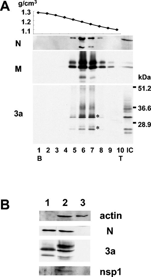
FIG. 4.
Western blot analysis of N, M, and 3a proteins in purified SCoV. Caco2 cells were infected with SCoV at a multiplicity of infection of 1, and culture fluid was collected at 5 days p.i. Released SCoV was purified by sucrose gradient centrifugation as described in the text. (A) Ten fractions from a 20 to 60% sucrose gradient containing the virus particles were collected and numbered from bottom (B) to top (T) of the gradient. The top panel represents the density of each sucrose fraction. IC, intracellular proteins from SCoV-infected Caco2 cells. (B) Purified SCoV (lane 1), cell extracts from SCoV-infected Caco2 cells at 5 days p.i. (lane 2), and uninfected Caco2 cells at 5 days p.i. (lane 3) were analyzed for actin protein, N protein, 3a protein, and SCoV nsp1 protein.

