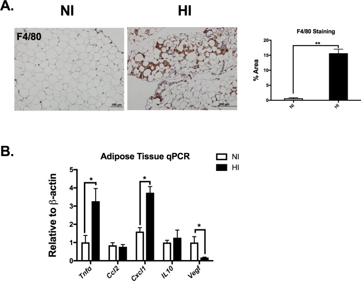
Fig 5. Iron deposition is associated with increased adipose tissue inflammation.
A) Representative adipose tissue F4/80 inmmunoflorescent staining from both NI (left) and HI (right) groups. Quantification for F4/80 antibody staining revealed macrophage clustering among the adipocytes in HI group. B) eAT gene expression with inflammatory gene markers. Abbreviations: Tnfα, tumor necrosis factor; Ccl2, C-C Motif Chemokine Ligand 2; Cxcl1, C-X-C Motif Chemokine Ligand 1, Il10, interlukin 10; Vegfα, Vascular endothelial growth factor A. *p< 0.05.

