
Figure 1.
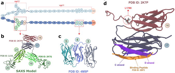
Schematic representation of Filamin. (a) Schematic representation of FLN dimer having 24 Ig-domains (blue) and the actin-binding domain (ABD, orange) in monomer. The two monomers are dimerized via domains 24. The atomic detailed structures of Ig-domains that form compact structures by interacting with the neighboring domains are highlighted and shown in green, and the domains 16 and 17 studied here are shown in light red and light blue, respectively. The structures of compact domain fragments are shown in panel (b) (FLNa16-2121) and in panel (c) (FLNa3-520). The FLNa16-21 model21 shown in (b) is obtained from SAXS using the high resolution structures of FLNa16-1722, FLNa18-1922, and FLNa20-2123. (d) Shows the structure of the domain pair FLNa16-1722 studied here. The binding mode of GPIbα-peptide (orange) to groove between the strands C and D of FLNa17 is shown. The FLNa16-17 - GPIbα-peptide structure is obtained by superimposing FLNa17-GPIbα-peptide X-ray structure26 with FLNa16-17 structure22. L1788, whose mutation to arginine causes FMD29 is shown as ticks.