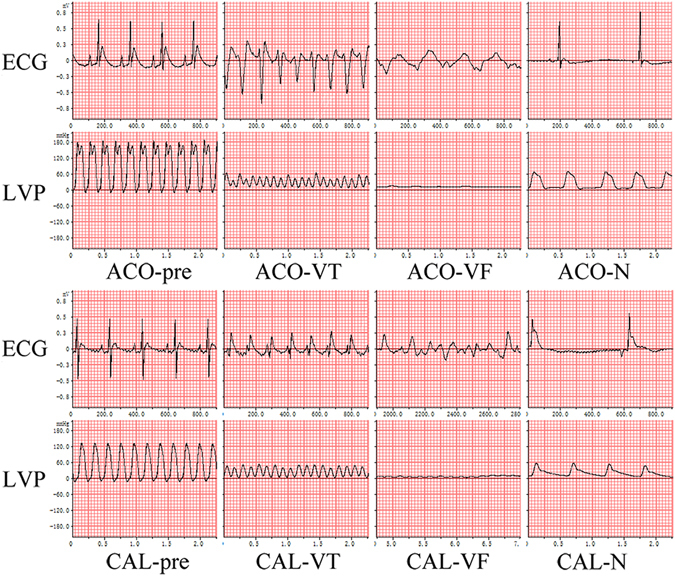
Figure 1.

ECG and hemodynamic features of two LVTA models and controls. Both LVTA models (ACO-VT, ACO-VF, CAL-VT, CAL-VF) experienced VT, VF, and a dramatic decrease in LVP. Control subjects (ACO-N and CAL-N) experienced bradycardia and mildly decreased LVP when compared to controls; ACO-pre and CAL-pre: measurements taken prior to treatments; measurements during VT or VF in LVTA models; ECG: electrocardiogram; LVP: left ventricular pressure; VT: ventricular tachycardia; VF: ventricular fibrillation.
