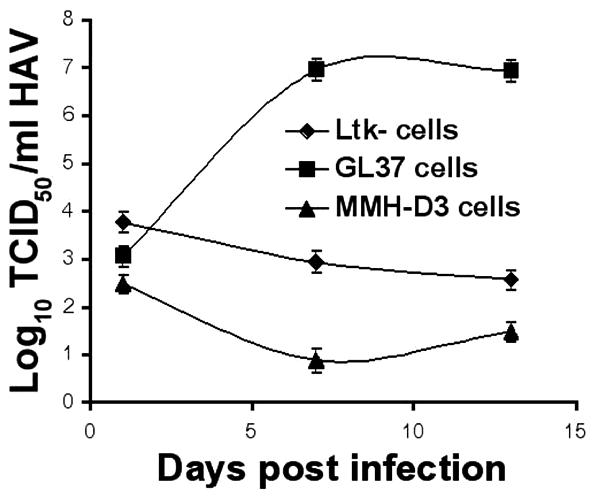
FIG. 1.

Growth of HAV in cell lines after infection. Monolayers of MMH-D3, Ltk-, and GL37 cells grown in 25-cm2 flasks were infected with HAV PI at an MOI of 1 to 2 TCID50/cell. At 24 h postinfection, cells were washed three times, split 1:3, and grown at 35°C in a CO2 incubator. Cells were split 1:3 weekly; approximately one-third of the collected cells were subjected to three freeze-thaw cycles, cell debris was pelleted, and the supernatant containing HAV was stored at −70°C. HAV was titrated by an endpoint ELISA in 96-well plates containing GL37 cell monolayers. Values are log10 HAV titers determined by the method of Reed and Müench (29). Error bars, standard deviations.
