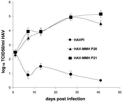
FIG. 5.
Growth of HAV-MMH in MMH-D3 cells cultivated under optimal conditions. Monolayers of MMH-D3 cells grown in six-well plates with a medium containing EGF and IGF II were infected with parental HAV PI, HAV-MMH P20, or HAV-MMH P21 at an MOI of 0.2 TCID50/cell. At 24 h postinfection, cells were washed three times and incubated at 35°C in a CO2 incubator. Cells were split 1:3 weekly; approximately one-third of the remaining cells were subjected to three freeze-thaw cycles, cellular debris was pelleted, and the HAV in the supernatant was titrated as described in the legend to Fig. 1. Error bars, standard deviations.

