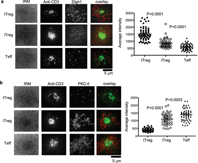
Figure 1.

Dlgh1 and PKC-θ are recruited differently to IS in tTregs, iTregs and Teffs. MACS bead purified human CD4+CD25hi (tTreg), CD4+CD25− T cells activated by TGF-β and Rapamycin (iTregs) and CD4+CD25− T cells (Teffs) were introduced into bilayers containing both anti-CD3 (5 µg/ml) and ICAM-1 at 250 molecules/mm2 fixed at 20 min and permeabilized, stained with anti-Dlgh1 (a) and anti-PKC-θ (b) antibodies and imaged by TIRFM. The panels show representative images. Dlgh1 and PKC-θ staining was quantified by calculation of average fluorescence intensity in cells. Data are representative of two different experiments. P values were calculated using Mann-Whitney test.
