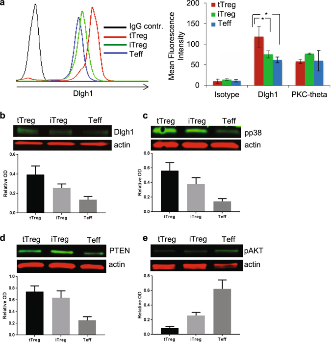
Figure 3.

Intracellular levels of Dlgh1 and PKC-θ were determined by intracellular staining by FACS (a) or by Western Blot (b) in tTregs, iTregs and Teffs. Cells were activated by immobilized anti-CD3 antibodies (5 μg/ml) and lysed. Phosphorylation of p38 (c) and AKT (d) as well as PTEN protein levels (e) was determined by Western Blot. Data are representative of 3 different experiments, the bar charts (B-E) are means ± SEM. *P < 0.05 was calculated by t test. Full-length gels are included in Supplementary Figure 2.
