
Figure 1.
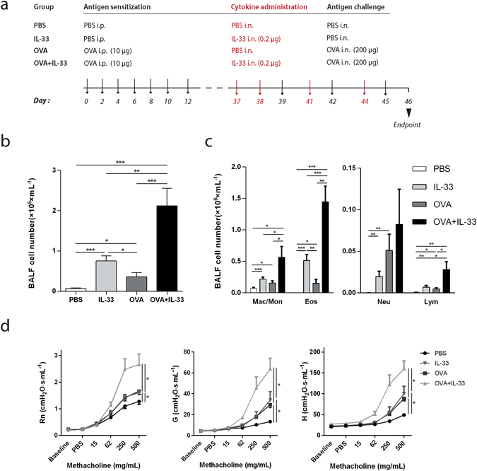
IL-33 potentiates antigen induced inflammation in BALF and airway hyperresponsiveness. (a) Schematic representation of the exposure protocol. Sham-or OVA-sensitized C57BL/6 mice received intranasal instillations of IL-33 or PBS. (b) Total inflammatory cells in BALF were counted. (c) Differential cell counts in BALF were determined. Results expressed as cells/mL for macrophages/monocytes, eosinophils, neutrophils and lymphocytes. (d) Airway hyperresponsiveness (AHR) in response to inhaled methacholine was measured in sham- or OVA-sensitized C57BL/6 mice that received intranasal instillations of IL-33 or PBS. Maximal responses to increasing doses of methacholine are shown for newtonian resistance (Rn), tissue damping (G) and tissue elastance (H) (b–d) *p < 0.05, **p < 0.01, ***p < 0.001 (ANOVA, Bonferroni). Results are pooled data from four independent experiments (mean ± SEM of n = 9–10 mice for each group).