FIG. 3.

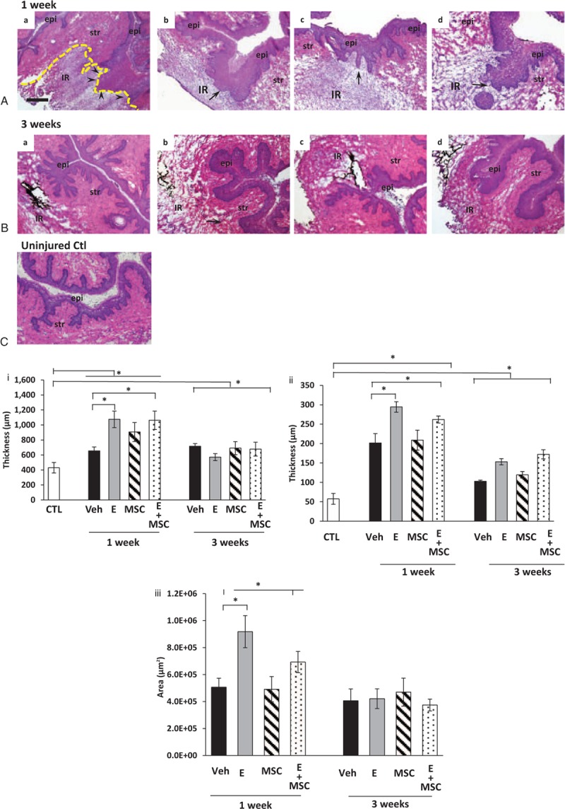
Histologic analysis of injury sites from ovariectomized rats treated with placebo, estrogen, MSCs, or MSCs + E at 1 (A) or 3 (B) weeks. epi, epithelium, str, stroma; IR, injury reaction. Arrows denote basement membrane at site of injury. Arrowheads in Aa indicate disruption of the basement membrane. (C) Uninjured control. Sections were stained with hematoxylin and eosin and captured at the same magnification (bar = 400 μm). Dark staining indicates incorporation of India ink at injury site. Dashed line indicates area of injury reaction. Lower panel (i), thickness of posterior vaginal stromal compartment or epithelium, (ii) at the injury site (μm) from ovariectomized rats treated with placebo cream/placebo injection (Placebo, solid bar), estrogen cream/placebo injection (E, gray bar, n = 9), placebo cream/mesenchymal stem cell injection (MSC, lined bar), or estrogen cream + MSC (E + MSC, dotted bar) 1 and 3 weeks after injury. Results were compared with uninjured untreated control ovariectomized rats (CTL). (iii) Area of injury reaction (μm2). Data represent mean ± SEM of 9-10 animals in each treatment group except uninjured controls (n = 3). ∗Two-way analysis of variance (P < 0.05). Veh, vehicle; CTL, control.
