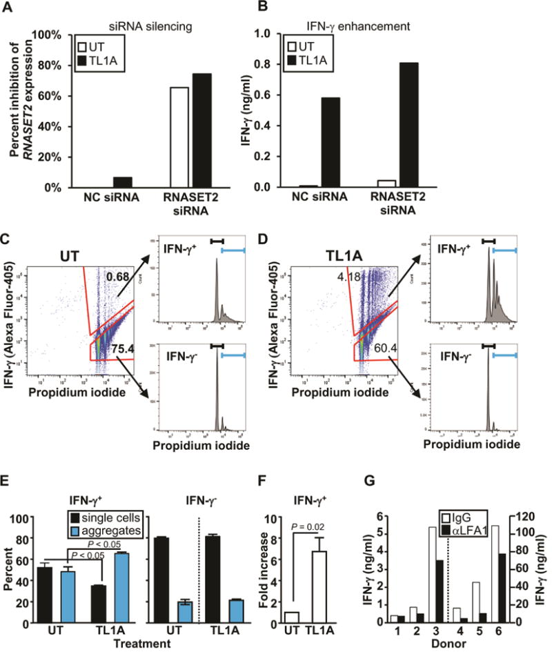
Fig. 6.

Effect of RNASET2 silencing on IFN-γ secretion and cellular Aggregation. (A) Silencing of RNASET2 expression by RNASET2 or control (NC) siRNA. (B) Effect of RNASET2 silencing on IFN-γ secretion. Panels A and B are representative of 6 out of 7 experiments (Fig. S7) with similar results. (C–D) CD4+ T cells were either (C) not treated (UT) or (D) stimulated with TL1A. Intracellular IFN-γ staining and cellular aggregation were measured by flow cytometry. Cells were gated on IFN-γ secreting and non-secreting populations (left panels) and then using propidium iodide (PI) analyzed for single and aggregate cell fractions (histograms, right panels). The first peak in each histogram corresponds to single cells (black bracket) and the remaining peaks to cellular aggregates (blue bracket). Representative of 4 experiments. (E) Proportion of single cells and cellular aggregates in IFN-γ secreting (IFN-γ+) and non-secreting (IFN-γ−) populations following TL1A stimulation. (F) Fold increase in number of IFN-γ secreting cells (average of 4 experiments). (G) CD4+ T cells were pretreated with control IgG or LFA1 blocking Ab (aLFA1) prior to TL1A stimulation. Overall p value for LFA-mediated blocking of IFN-γ secretion, measured by ELISA, was 0.047.
