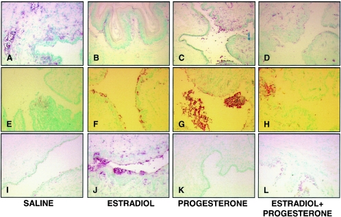
FIG. 7.
Localization of neutrophils in vaginal tissue of OVX, hormone-treated mice infected with HSV-2. A rat anti-mouse neutrophil antibody was used to detect specific staining, as described in Materials and Methods. Mice were sacrificed either 24 h postinfection (A to D) or 3 days postinfection (E to H). Control noninfected mice that received hormones were also examined on the same day as the mice examined 3 days postinfection (I to L). Positive staining (pink) is seen in the endothelium of saline-treated mice on day 1 (A) and mostly following infection of progesterone-treated mice (C and G). Significant numbers of neutrophils are also seen in the superficial layers of vaginal epithelium 3 days after E2 treatment was stopped in both infected and noninfected tissue (F and J). Original magnification, ×100.

