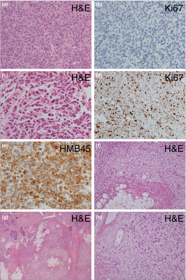
Figure 2.

Congenital melanocytic naevus (CMN) – histological features in the nervous system (a–e) and skin (f–h). (a, b) Images of leptomeningeal disease showing a cellular collection of melanocytes with minimal atypia and no significant proliferation, confirmed on Ki67 labelling (b) (patient 3, Table 1). (c–e) In contrast, proliferation of markedly atypical cells with frequent mitotic figures and a high Ki67 labelling index (e). The lesion expresses markers of melanocytes (HMB45). (f–h) Areas in a proliferative nodule within a cutaneous CMN demonstrating typical small deep melanocytes admixed with expansile areas formed of spindled cells and areas with larger cells with eosinophilic cytoplasm; there is no significant atypia and no mitoses are seen. H&E, haematoxylin and eosin.
