
Figure 3.
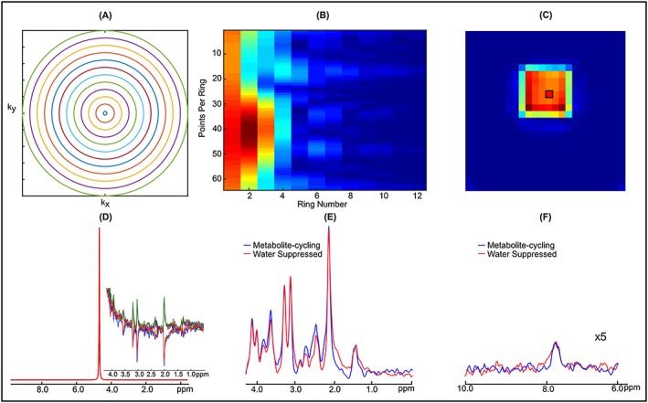
A, Concentric ring k‐space trajectory used for the braino phantom. B, k‐space data acquired using non‐water suppressed metabolite‐cycling MRSI with following parameters: N ring = 12, N p_ring = 64, FOV = 240 mm × 240 mm, STEAM localization =65 mm × 65 mm × 20 mm, T R = 2 s, T E = 14 ms, T M = 32 ms, ADC bandwidth =80 kHz, N avg = 10 and maximum slew rate = 67.8 mT/m/ms. C, Water image with a final grid of 24 × 24 (2N ring × 2N ring) obtained using the first time point of the water FID. D, Spectra of 10 non‐water‐suppressed water peaks from a voxel taken from the STEAM localized region (black box in C). The subfigure illustrates the effect of asymmetric RF pulses on the upfield spectrum. E,F, Representations of upfield (E) and downfield (F) spectra extracted from a 2 mL voxel (black box in C) from the data acquired using non‐water‐suppressed (blue) and water‐suppressed (red) STEAM localization. The residual water peak was filtered with the HLSVD algorithm. Phantom spectra were line broadened (6 Hz) to match line widths encountered in vivo