Figure 2.

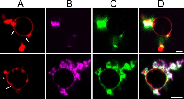
Fibrillization‐induced membrane deformations: POPC/POPG GUVs doped with Rh‐DOPE were incubated with 20 μm (equivalent monomer concentration) ThT labelled fibril seeds and 80 μm αS monomers (1 mol % αS‐AlexaFluor647) at 37°C in quiescent conditions. Images were taken 3 hours after the start of the experiment. A) Rh‐DOPE fluorescence (red). B) αS‐Alexa647 fluorescence (magenta). C) ThT fluorescence (green). D) Overlay of A, B and C. The addition of αS to fibril seeds results in the growth of ThT positive amyloid fibrils from the seeds at both the membrane and in solution. The white arrows indicate some of the places where fibril growth has resulted in deviations from the normally nearly spherical vesicle shape. Scale bar: 5 μm.
