Figure 3.

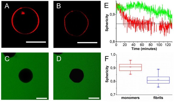
Fibrillization‐induced deformation of POPC/POPG GUVs: A) An initially spherical vesicle is incubated with 95 μm αS monomers and 5 μm fibril seeds B) as a result of fibril growth it deforms and becomes a polyhedron (Δt=4 hours). When POPC/POPG vesicles were incubated with 20 μm αS seeds and 80 μm αS monomers no influx of calcein dye (green) could be observed. C) Immobilized vesicle at t=0 and D) t=160 minutes after the start of the experiment. E) To quantify the shape changes the sphericity of the vesicles was followed in time. The black line indicates the sphericity of an icosahedron, the green and red line follows the shape changes of the GUVs shown in A, B and C, D respectively. E) Sphericity of POPC:POPG 1:1 GUVs in the presence of αS monomers (red) and after incubation with growing αS fibrils. The average sphericity (open squares), the mean sphericity (middle line) and the 25 and 75 percentile (box) are shown. All scale bars: 10 μm.
