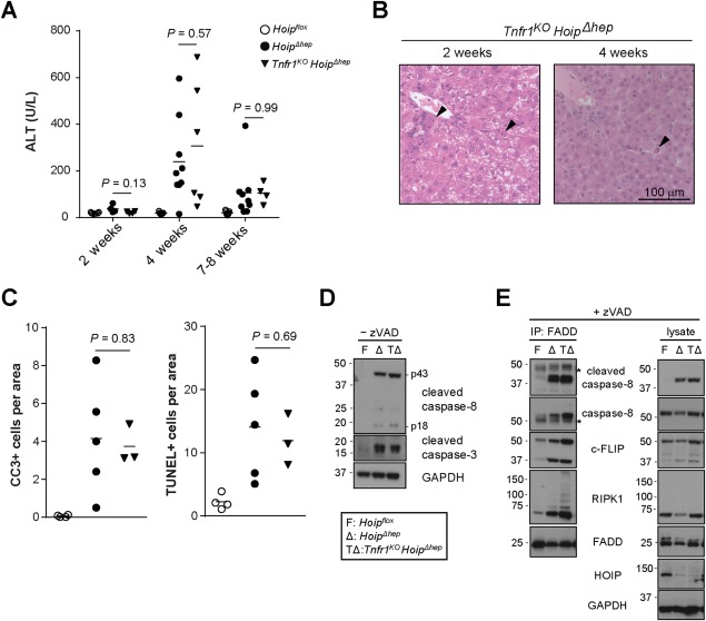
Figure 6.

Hepatocyte apoptosis in HOIP‐deficient livers is unaffected by genetic ablation of TNFR1. (A) Levels of serum ALT in Hoipflox, HoipΔhep, and Tnfr1KO HoipΔhep mice at the indicated ages. (B) Representative pictures of hematoxylin and eosin sections of the livers of Tnfr1KO HoipΔhep mice at 2 and 4 weeks of age. Arrowheads indicate apoptotic bodies. (C) Quantification of CC3+ and TUNEL+ cells per area of the livers of Hoipflox, HoipΔhep, and Tnfr1KO HoipΔhep mice at 2 weeks of age. (D) Lysates of primary hepatocytes from Hoipflox (F), HoipΔhep (Δ), and Tnfr1KO HoipΔhep (TΔ) mice at 8 weeks of age were immunoblotted for CC8 and CC3. (E) Immunoprecipitation of the FADD‐associated complex in zVAD‐treated (24 hours) primary hepatocytes from Hoipflox (F), HoipΔhep (Δ), and Tnfr1KO HoipΔhep (TΔ) mice. Asterisks indicate unspecific bands. Unpaired two‐tailed t test was employed for statistical analysis. Abbreviations: GAPDH, glyceraldehyde 3‐phosphate dehydrogenase; IP, immunoprecipitation.
