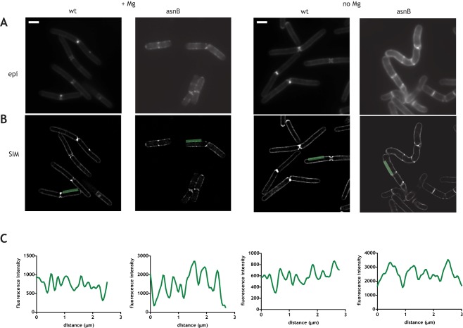
Figure 5.

Isotropic insertion of PG along the sidewalls of the wild‐type and ΔasnB mutant cells. Epifluorescence (A) and structured illumination microscopy (B) imaging of insertion of peptidoglycan by TDL labeling in the presence of added Mg2+ (two panels on the left) and in the absence of added Mg2+ (two panels on the right). Structured illumination microscopy (SIM) images of the medial plane of the cell are shown. C. Quantification of fluorescence along the green line shown in SIM images. For SIM images of additional cells see Supporting Information Fig. S6.
