
Figure 1.
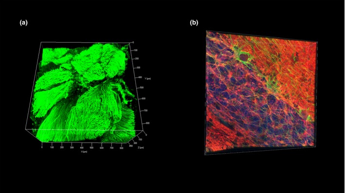
Human brain tissue processed and immunostained using free of acrylamide sodium dodecyl sulphate (SDS)‐based tissue clearing (FASTClear). (a) z‐Stack image of a ventral root of a piece of spinal cord fresh tissue (3 mm thick) immunostained using antineurofilament primary antibody (final concentration 1:100; Dako M0762) and Alexa‐fluor 488 conjugated donkey anti‐mouse secondary antibody. Stained tissue was visualized using a Zeiss 780 inverted confocal microscope (Carl Zeiss, Germany) with ×10 objective (imaging depth to 508.519 μm, z‐stack step size 3.03 μm). (b) z‐Stack image of a piece of fixed cerebellar tissue immunostained using antibodies against neurofilament (green; final concentration 1:100; Dako M0762) and βIII‐tubulin (red; final concentration 1:100; Millipore AB9354) and counterstained with 4′,6‐diamidino‐2‐phenylindole (blue). Stained tissue was visualized using a Leica SP5 (Leica Microsystems, UK) confocal microscope with ×40 objective (imaging depth to 66.5 μm, z‐stack step size 0.38 μm).