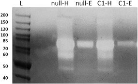
Figure 5.

Comassie blue stained casein Zymogram. From left: null HCCF (null‐H), null AIEX Eluate (null‐E), Cell line1 HCCF (C1‐H), Cell line 1 AIEX Eluate (C1‐E).
Protein samples are first separated according to molecular weight via gel electrophoresis, then renatured and incubated overnight in the gel containing casein substrate and finally stained and de‐stained. Areas that show proteolytic activity appear as white bands over dark background. Data show that AIEX is able to clear out the majority of proteases from HCCF to Eluate sample except for some co‐eluting ones at around 80 KDa. These were later putatively identified via MS as Dipeptidyl peptidase 3 and Prolyl endopeptidase. Furthermore, amount of protease in GAA cell line 1 eluate seems to be less than amount of proteases present in null eluate. Note: GAA, not being a protease, is not visible in zymography.
