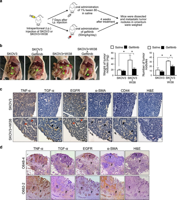
Figure 6.
Stromal fibroblasts promote peritoneal metastasis of ovarian cancer in vivo through activation of EGFR, and a TNFα-TGFα-EGFR interacting loop presents in the microenvironment of omental metastases of ovarian cancer. (a) Schematic diagram showing the use of orthotopic ovarian cancer xenograft model to evaluate the effect of stromal fibroblasts on peritoneal metastasis of ovarian cancer in vivo, and the effect of EGFR activation on fibroblast-induced peritoneal metastasis of ovarian cancer in vivo. (b) Macroscopical morphology of metastatic tumor nodules in orthotopic xenografts was shown (green arrow and green stroke). The weight and number of dissected metastatic tumor nodules were quantified (right). The weight and number of metastatic tumor nodules were compared between the saline-treated mice injected SKOV3+WI38 (n=5) and the saline-treated mice injected with SKOV3 alone (n=5), and also between the Gefitinib-treated mice injected with SKOV3+WI38 (n=5) and the saline-treated mice injected with SKOV3+WI38 (n=5). *P<0.05 (Student's t test). (c) Immunohistochemical staining for TNF-α, TGF-α, EGFR, CD44 and α-SMA in serial sections of metastatic tumor nodules collected from the saline-treated mice injected with SKOV3 alone or with SKOV3+WI38. Fibroblasts were identified by positive signal of α-SMA and CD44 staining. Image was captured at 200x magnification. (Red arrow: cancer cells and black arrow: fibroblasts) (d) Immunohistochemical staining for TNF-α, TGF-α, EGFR, and α-SMA in serial sections of omental metastases from patients with advanced ovarian cancer. Image was captured at 200x magnification. (T: ovarian tumor cells and F: omental stromal fibroblasts).

