Figure 1.
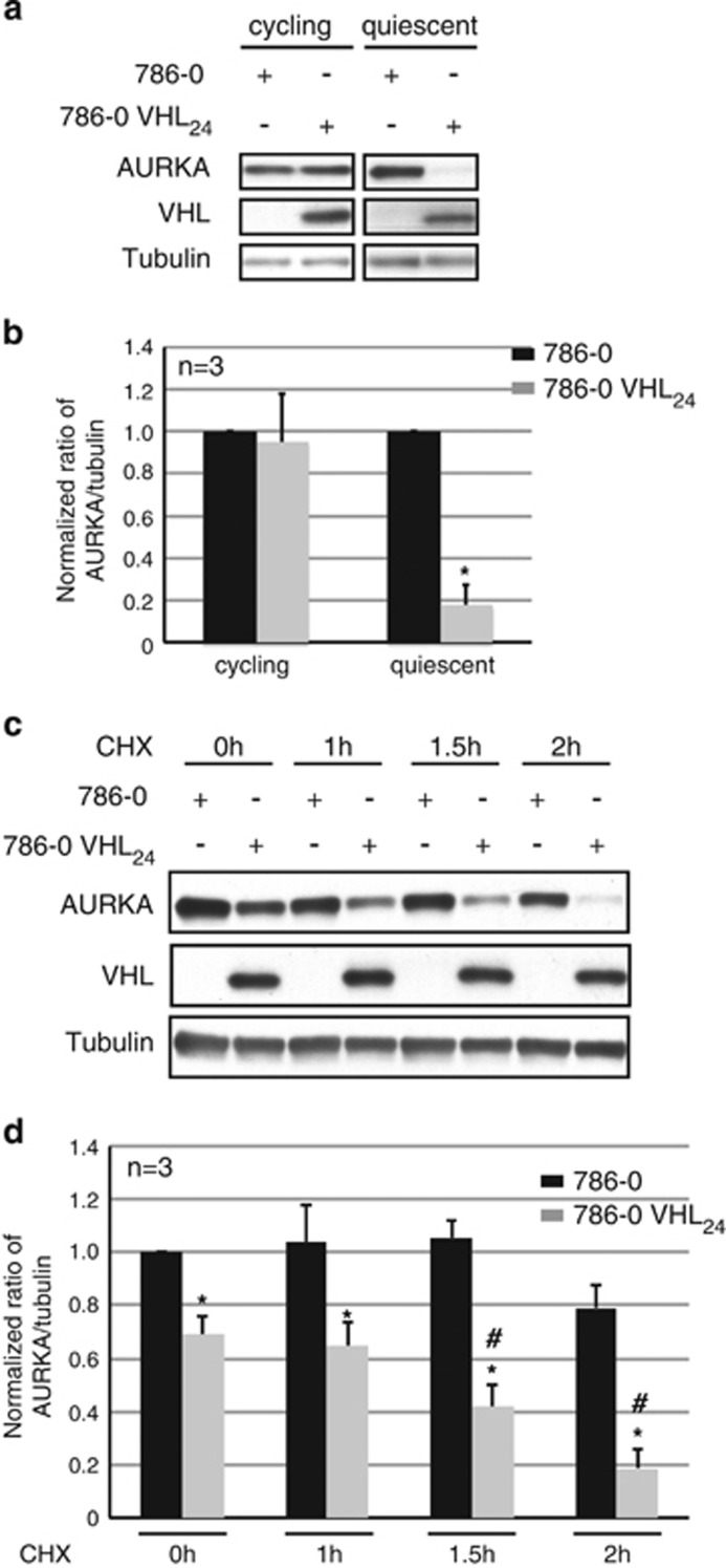
VHL modulates AURKA protein levels. (a) Lysates from 786-0 to isogenic 786-0 cells overexpressing VHL (VHL24) cultured at sub-confluent (cycling) or confluent and serum starved conditions (48 h) probed as indicated. (b) Densitometric quantitation of the average ratio of AURKA to tubulin expression from 786-0 (black bars) to 786-0 VHL24 (gray bars) cells. *P<0.01. (c) Lysates harvested from 786-0 to 786-0 VHL24 cells cultured to confluence and serum starved for 24 h before treatment with CHX for the indicated time points probed as shown. (d) Densitometric quantitation showing an average ratio of AURKA to tubulin in 786-0 (black bars) and 786-0 VHL24 (gray bars) cells treated with CHX. *P<0.01. # Denotes significant differences exclusively in the 786-0 VHL24 cell line at the each of the indicated CHX treatment time points compared with the 0- h time point (P<0.05). Error bars denote s.e.m.

