Figure 6.
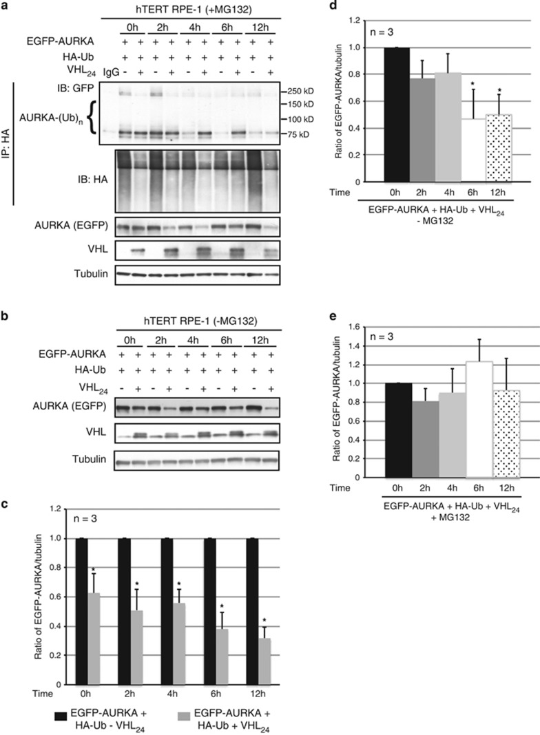
VHL promotes AURKA degradation as cells enter quiescence. (a) In vivo ubiquitination assay using hTERT RPE-1 cells overexpressing EGFP-AURKA and HA-Ub with and without overexpressed VHL24 in the presence of MG132, harvested following serum withdrawal at the indicated time points. Ubiquitinated AURKA immunoprecipitated using an anti-HA antibody and probed with an anti-AURKA antibody. Input lysates immunoblotted for the indicated antibodies. (b) Lysates from hTERT RPE-1 cells overexpressing EGFP-AURKA and HA-Ub with and without overexpressed VHL24 in the absence of MG132 probed for the indicated antibodies. (c) Densitometric quantitation showing an average ratio of EGFP-AURKA to tubulin in cells without (black bars) and with (gray bars) overexpressed VHL24 at the indicated time points. *P<0.01. (d, e) Densitometric quantitation of EGFP-AURKA to tubulin expression averaged from three independent experiments at each of the indicated time points normalized to the 0-h time point in the absence of MG132 treatment (d) and the presence of MG132 treatment (e). *P<0.01 (compared with the 0-h time point). All error bars denote s.e.m.

