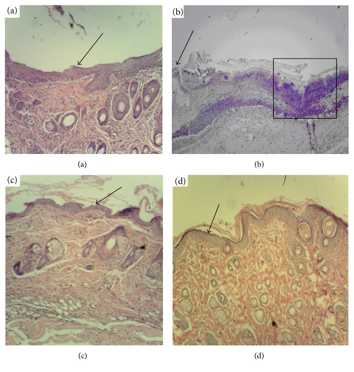
Figure 4.
Histological analysis of derma within the wound bed, day 10 after the beginning of curing (magnif. ×70). (a) Positive control (clotrimazole); (b) negative control (no specific treatment); (c) reference group (wild-type K. pinnata extract); (d) experimental group (CecP1). Selected area: arrow—new-formed thin epithelium, square—area of melting of the necrotic tissue.

