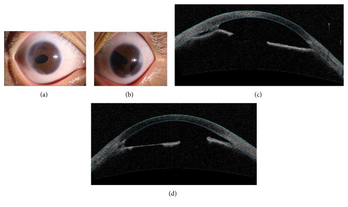
Figure 3.
Ocular characteristics of subject III-1. Anterior segment photography showed corectopia and peripheral anterior synechia in the right eye (a) and iris hypoplasia, corectopia, polycoria, and peripheral anterior synechia in the left eye (b). Anterior segment OCT showed iridocorneal adhesions in the right eye (c) and iris hypoplasia and iridocorneal adhesions in the left eye (d).

