Figure 6.

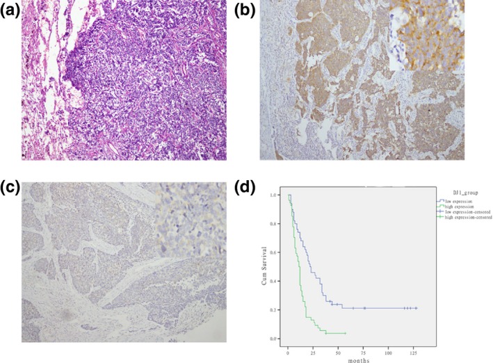
Expression of DJ‐1 in SCLC samples and the survival rates of SCLC patients. (a) HE staining of SCLC samples (magnification, ×100). (b,c) Representative images of immunostaining of DJ‐1 in small‐cell lung cancer tissues with DJ‐1‐high expression (b) and DJ‐1‐low expression (c) (magnification, ×50–400). (d) Kaplan–Meier analysis of correlation between DJ‐1 expression and the survival time of patients with SCLC (P < 0.001).
