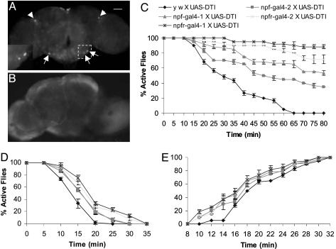
Fig. 2.
NPFR1 neurons mediate ethanol response. GFP expression was detected in the brain of flies from the npfr1-gal4 X UAS-GFP crosses. (A) Arrowheads indicate two dorsolateral neurons in the central complex and three representative neurons in the subesophageal ganglia. (A Inset) Magnified view of the boxed area. (B) However, this GFP expression pattern was abolished in the brain of npfr1-gal4 X UAS-GFP/UAS-DTI flies. Control flies (UAS-GFP without a gal4 driver) showed no such GFP expression pattern in the brain (data not shown). (C) The alcohol sensitivity of experimental and control flies was determined under the same conditions as above. In three separate trials (n = 20 per trial), the significantly higher percentages of active flies were consistently observed for npfr1-gal4-1 and -2 X UAS-DTI flies than for npf-gal4-1 and -2 X UAS-DTI flies. The y w X UAS-DTI controls showed least ethanol resistance (P < 10-4, starting from 30 min). The flies were also tested for ethanol resistance under the same conditions, except a higher dose (1 ml of 86% ethanol) was used (D, P < 0.001, starting from 15 min). (E) To measure the rate of recovery from ethanol anesthetization, flies were exposed to concentrated ethanol vapor for 12 min, allowed to recover under the ethanol-free conditions, and the number of flies capable of climbing after the ethanol treatment was counted. The data of fly recovery over the time interval of 8-14 min after ethanol treatment were analyzed statistically. The control fly (y w X UAS-DTI) showed a delay in restoring normal locomotion compared with the four experimental flies (P < 10-4).

