Figure 5.

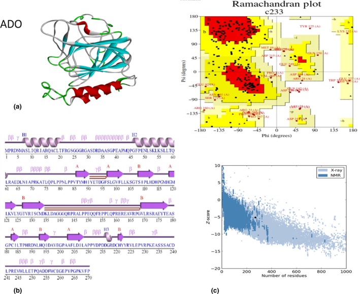
(a) Modelled structure of cysteamine dioxygenase (ADO) [SWISS‐MODEL server (Laskowski, 2009)]; (b) 2‐D structure of ADO from PDBsum is modelled from SWISS‐MODEL modeling software; and (c) PROCHECK analysis and Ramachandran plot of the 3‐D structure of ADO. The protein model has 83.6% of amino acids in the most favored region, 11.7% of amino acids as additional allowed region, 2.9% of amino acids generously allowed region, and 1.8% amino acids as disallowed region. Protein structure analysis (Pro SA‐web) predicted that ADO protein has Z‐score of −5.79.
