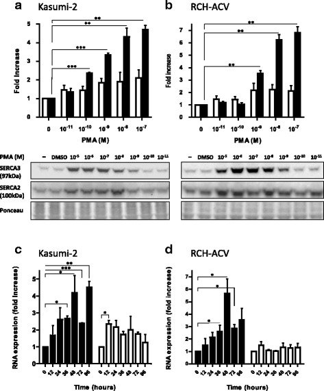
Fig. 1.

Induction of SERCA3 expression in precursor B ALL cell lines. Kasumi-2 (a) and RCH-ACV (b) cells were treated by various concentrations of PMA for 5 days, and SERCA3 (closed columns; 97 kDa) as well as SERCA2 (open columns; 100 kDa) expression was detected by Western immunoblotting (lower part) and quantified (upper part). Equal total cellular protein loading of blots was checked by Ponceau red staining. PMA treatment led to an approximately five- or sevenfold increase of SERCA3 expression in Kasumi-2 and RCH-ACV cells, respectively. Concentration-dependent induction of SERCA expression by PMA could be observed from 10−10-10−9 M and reached a plateau around 10−8-10−7 M, whereas DMSO vehicle was without effect. c and d: Kasumi-2 and RCH-ACV cells were treated with 10−8 M PMA, and SERCA3 (closed columns), as well as SERCA2 mRNA levels (open columns) were measured by quantitative RT-PCR and normalized to GAPDH as described in Methods. When compared to untreated cells, SERCA3 mRNA expression peaked at day 2 of treatment, and a rebound could be observed at day 4
