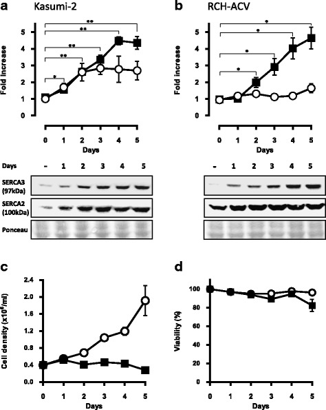
Fig. 2.

Time course of SERCA expression in PMA-treated precursor B ALL cell lines. Kasumi-2 (a) and RCH-ACV (b) cells were treated with 10−7 M PMA and SERCA3 (closed squares) as well as SERCA2 (open circles) expression was detected by Western blotting (lower part) and quantified (upper part). Equal protein loading was checked by Ponceau red staining of the membranes. Induction of SERCA3 expression was detected from days 1-2 and reached a plateau after 4-5 days. Treatment (Kasumi-2 cells, PMA: 10−8 M) led to rapid growth arrest (c) with maintained viability (d) during 5 days
