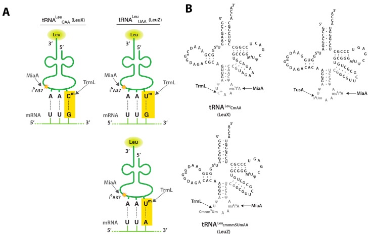
Figure 1.
tRNA (cytidine/uridine-2’O)-ribose methyltransferase L (TrmL), tRNA dimethylallyltransferase (MiaA), and tRNA 2-thiouridine synthesizing protein A (TusA) tRNA modifications and UUX-Leu codon recognition; (A) Schematic depicting tRNALeuCAA (LeuX) and tRNALeuUAA (LeuZ) anticodons and their cognate mRNA codons (UUG or UUA). This schematic is modified from a leucine tRNA schematic obtained from the following source [20]; (B) tRNALeuCAA (LeuX) and tRNALeuUAA (LeuZ) secondary structure secondary structure. Nucleotides subject to modification are shown in grey and the sites of the MiaA, TrmL, and TusA modifications studied here are shown. This schematic is modified from Figure 3C of [21].

