Fig. 6.

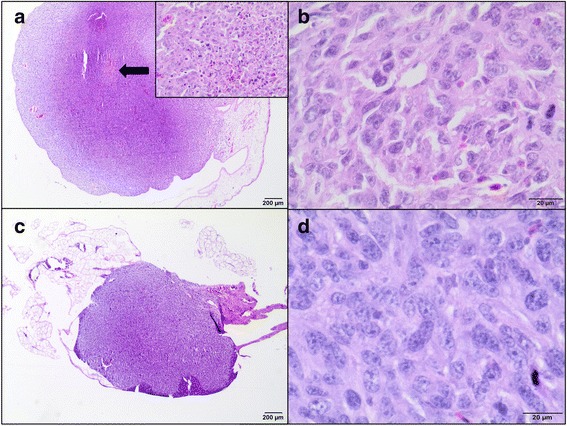
Hematoxillin and eosin staining of tumors growth from feline fibrosarcoma FFS1 and FFS3 cell lines on the CAM. a Unencapsulated, well-demarcated tumor mass of feline sarcoma FFS1 cell line on the CAM. Neoplastic cells demonstrate an invasive growth pattern and are able to penetrate the CAM. An area of necrosis is indicated by arrow. An insert image shows more details of necrosis: neoplastic cells undergo nuclear pycnosis and karyorrhexis. H&E, magnification 20× (200× insert). b Higher magnification of tumor derived from FFS1 cell line. Densely cellular neoplasm composed of polygonal cells with indinstinct cell borders on a fine fibrovascular stroma. Neoplastic cells have a small to moderate amount of pale eosinophilic cytoplasm, round to elongate nucleus with variably distinct magenta nucleolus. Nuclear pleomorphism is marked. Mitotic figures average 2-4 per HPF with often bizarre mitoses. H&E, magnification 400×. c Tumor growth of feline sarcoma FFS3 cell line on the chick embryo chorioallantoic membrane, well-circumscribed, unencapsulated and invaded CAM. H&E, magnification 20×. d Higher magnification of tumor derived from FFS3 cell line. Dense pleomorphic cell population composed of neoplastic cells with indistinct cell borders supported by a fine fibrovascular stroma. Neoplastic cells have pale eosinophilic cytoplasm, an oval to elongate nucleus with finely stippled chromatin and a variably distinct magenta nucleolus (up to 2). The mitotic rate is up to 1-2 per HPF. H&E, magnification 400×
