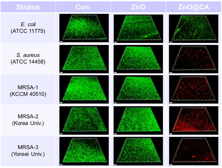
Figure 6.
Live and dead cell images of bacteria (E. coli, S. aureus, MRSA-1, MRSA-2, and MRSA-3) using a confocal fluorescence microscope after incubation with ZnO and ZnO@CA nanoparticles. The bacterial cells of the five strains on the cover glass were incubated at 35 °C with ZnO and ZnO@CA nanoparticles for 24 h. The images show live and dead bacterial cells stained with SYTO-9 (green) and propidium iodide (PI, red) fluorescent dyes, respectively. All samples were tested in duplicate for each experiment, and each experiment was repeated three times (n = 6). There were no significant differences on live and dead cell imaging in each sample. Scale bars represent 50 μm.

