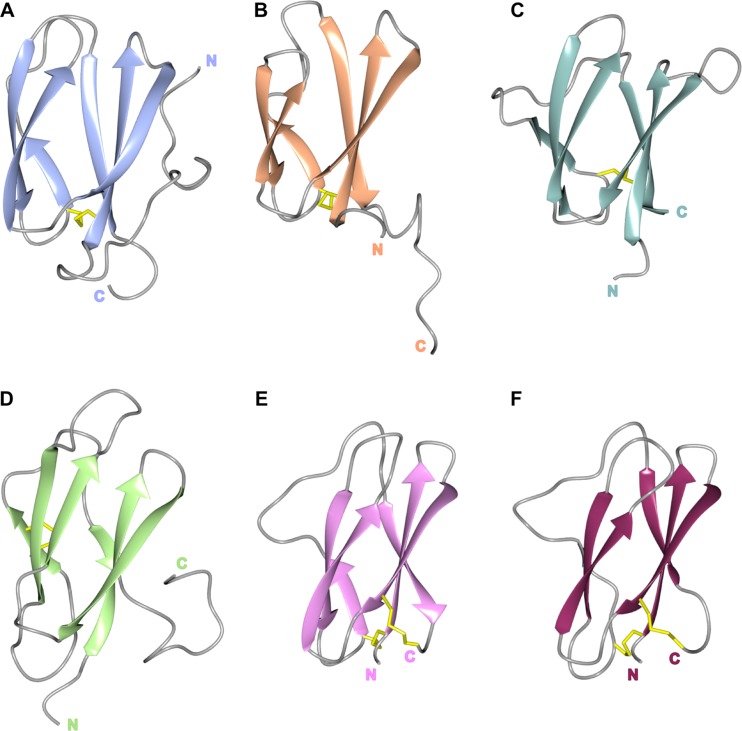
FIG 5.
The structures of MAX effectors reveal the shared β-sandwich fold. The conserved β-strands are shown in a cartoon representation for each protein, with residues contributing to disulfide bridges shown as sticks (in yellow) and loops in gray. Shown are AVR-PikD (A), AVR1-CO39 (B), AVR-Pia (C), AVR-Pizt (D), ToxB (E), and toxb (F), with amino (N) and carboxyl (C) termini labeled.

