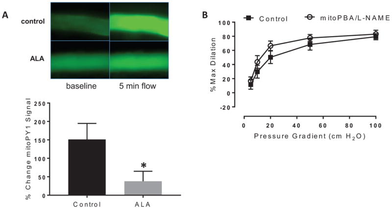
Figure 3.

Source of H2O2 after PGC-1α overexpression on flow-mediated dilation (FMD) in CAD vessels. A) Reduction in MitoPY1 fluorescence following 24-hour ALA treatment (250 μM). Changes in fluorescence intensity in response to shear stress were evaluated in untreated or ALA-treated CAD vessels. n=6-7 per treatment group. *P < 0.05 ALA vs control; B) mitoPBA had no additional effect on dilation after incubation with the eNOS inhibitor L-NAME following 24-hour ALA treatment (250 μM) in CAD vessels. P=NS vs control between flow curves.
